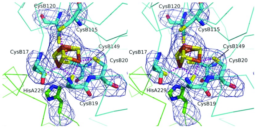
Figure 4. The large subunit His229 is close to the proximal cluster of the small subunit.
Stereoview of the [4Fe–3S] proximal cluster in Hyd-5. The associated cysteine residue ligands, and His229, are shown with the omit difference density map. The residues marked A originate in the α-subunit (large subunit), and those marked B originate in the β-subunit (small subunit). The map (blue chicken wire) is contoured at the 4σ level. The S positions are coloured yellow, Fe3+ brown, N blue and O red. The C positions of the small subunit are cyan and the large subunit green. Broken lines represent co-ordination links between S and Fe3+.

