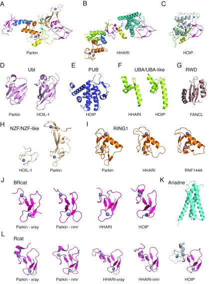
Figure 3. Catalogue of three-dimensional structures for RBR E3 ubiquitin ligases.
The upper panels show cartoon representations of multi-domain structures for (A) RING0–RBR from human parkin (PDB code 4I1F [35]; also PDB code 4K7D [34] and PDB code 4BM9 [36]), (B) human HHARI (PDB code 4KBL [42]) and (C) C-terminus of human HOIP (PDB code 4LJP [43]). The lower panels (D–L) show cartoon diagrams of three-dimensional structures of the individual domains for (D) Ubl domains from parkin (PDB code 2ZEQ [136]) and HOIL-1 (PDB code 2LGY [81]), (E) PUB domain from HOIP (PDB code 4JUY), (F) UBA-like domains from HHARI (PDB code 4KBL [42]) and HOIP (PDB code 4DBG [32]), (G) RWD from the E3 ligase FANCL (PDB code 3K1L [108]), (H) NZF and double NZF-like domains from HOIL-1 (PDB code 3B0A [39]) and parkin (PDB code 4I1F [35]), (I) RING1 domains from parkin (PDB code 4I1F [35]), HHARI (PDB code 4KBL [42]) and RNF144A (PDB code 1WIM), (J) BRcat domains from parkin (PDB code 4I1F [35] and PDB 2JMO [116]), HHARI (PDB code 4KBL [42]) and HOIP (PDB code 2CT7), (K) Ariadne domain from HHARI (PDB code 4KBL [42]), and (L) Rcat domains from parkin (PDB code 4I1F [35] and PDB code 2LWR [48]), HHARI (PDB code 4KBL [42] and PDB 2M9Y [117]) and HOIP (PDB code 4LJP [43]). The colour scheme for each individual domain and multidomain structures are as shown in Figure 2. Representative secondary structures are also labelled.

