
Figure 6. Evidence for flexibility in the RBR E3 ubiquitin ligases.
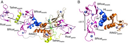
(A) The three-dimensional structures of parkin (PDB code 4I1F [35]) and HHARI (PDB code 4KBL [42]) are shown following superposition of their RING1 domains. This presentation shows the Rcat domains for the two proteins are found at opposite ends of the respective structures with respect to the location of the RING1 domain with large distances between the RING1 and Rcat domains (~32 Å in parkin and ~67 Å in HHARI) that must somehow be bridged for ubiquitin transfer. Regions not modelled in the parkin structure, probably due to flexibility, are indicated by broken lines. For clarity the UBA-like and Ariadne domains from HHARI are not shown. (B) The position of the BRcat domains in parkin and HHARI are shown after superposition of the RING1 domains. The position of the BRcat domain deviates ~22 Å between the parkin and HHARI structures due to an approximate 90° difference in the tilt of the RING1–BRcat interdomain helix. Only the RING1 domain from parkin is shown for clarity. The colour schemes used in (A) and (B) are as described previously in Figures 2 and 3.