
Figure 2.
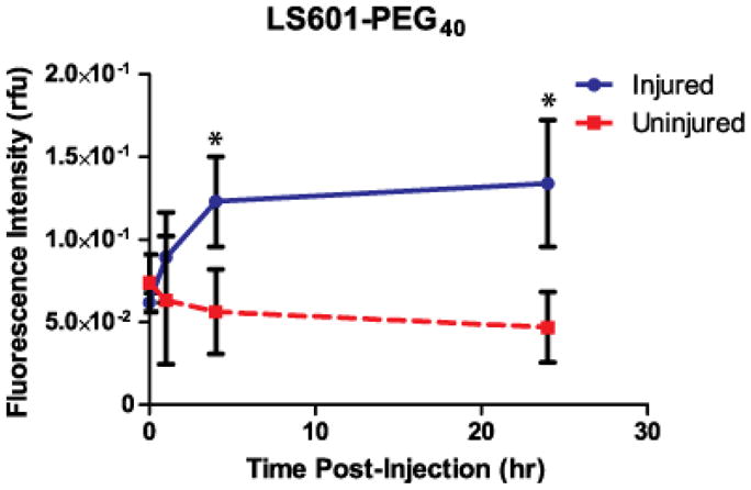
Fluorescence intensity (in relative fluorescence units) for ROIs selected from the lower limbs of mice at given timepoints after intravenous injection of LS601-PEG40 (n=3 mice). Based upon our results (see Results section), which indicate the free dye clears from injured control animals within 4 hours, attachment to PEG40 does result in significantly higher circulation times within which detection of ROS could take place. The fluorescence intensity increased about 2-fold in the injured limb over 24 hr. Bars indicate standard deviation and (*) indicates statistically significant (P<0.01) differences.