
Figure 6.
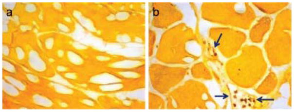
Immunohistochemistry of thigh muscle tissue for nitrotyrosine. Positive signal is brown and the counterstained tissue is yellow-orange. (a) Control thigh muscle shows little evidence of nitrotyrosine. (b) Ischemic thigh muscle shows positive staining in inflammatory cells (arrows) within and around small intramuscular blood vessels. Results are representative for healthy and ischemic tissues from n=4 subjects, including those represented in Figure 4. Additional images are shown in Figure S2.