
Figure 4.
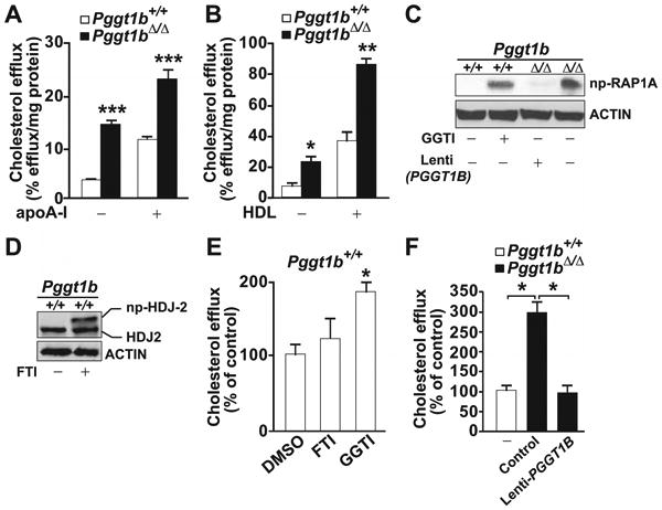
Increased cholesterol efflux in macrophages from Pggt1b∆/∆ mice. A and B, Basal and (A) apolipoprotein A1 (apoA1)– and (B) high-density lipoprotein (HDL)–stimulated cholesterol efflux in bone marrow (BM) macrophages (n=4 per genotype). C, Western blots demonstrating high levels of nonprenylated (np) RAP1A in Pggt1b∆/∆ BM macrophages and in Pggt1b+/+ macrophages incubated with a geranylgeranyltransferase type I (GGTase-I; 10 μmol/L) and low levels in Pggt1b+/+ cells and in Pggt1b∆/∆ cells incubated with a lentivirus expressing human PGGT1B. D, Control Western blots demonstrating reduced electrophoretic mobility of HDJ2 in Pggt1b+/+ cells incubated with a farnesyltransferase inhibitor (FTI; 10 μmol/L). E and F, Basal cholesterol efflux in BM macrophages incubated with dimethyl sulfoxide (DMSO), FTI, or GGTI (n=3–4 per treatment; E) or with lenti-PGGT1B at a multiplicity of infection of 20 (n=3; F). *P<0.05; **P<0.01; ***P<0.001.