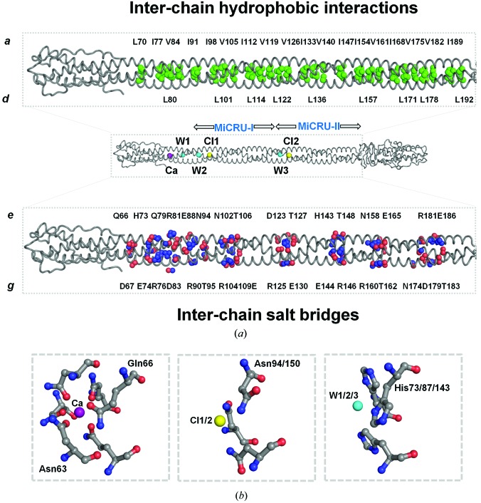
Figure 4.
Determinants for the structural stability of the gp26-2M fiber. (a) Middle: a cartoon (tube) representation of gp26-2M displaying buried ions and water molecules inside the helical core. Top: a magnified view of the gp26-2M helical core with the aliphatic side chains of β-branched chain hydrophobic residues shown as green spheres. Bottom: magnified view of the gp26-2M helical core showing charged residues involved in interchain salt bridges (O and N atoms are shown as red and blue spheres, respectively). (b) Magnified view of the calcium (Ca) ion, chloride ions (Cl1 and Cl2) and water molecules (W1, W2 and W3) buried inside gp26-2M cavities showing interacting residues.

