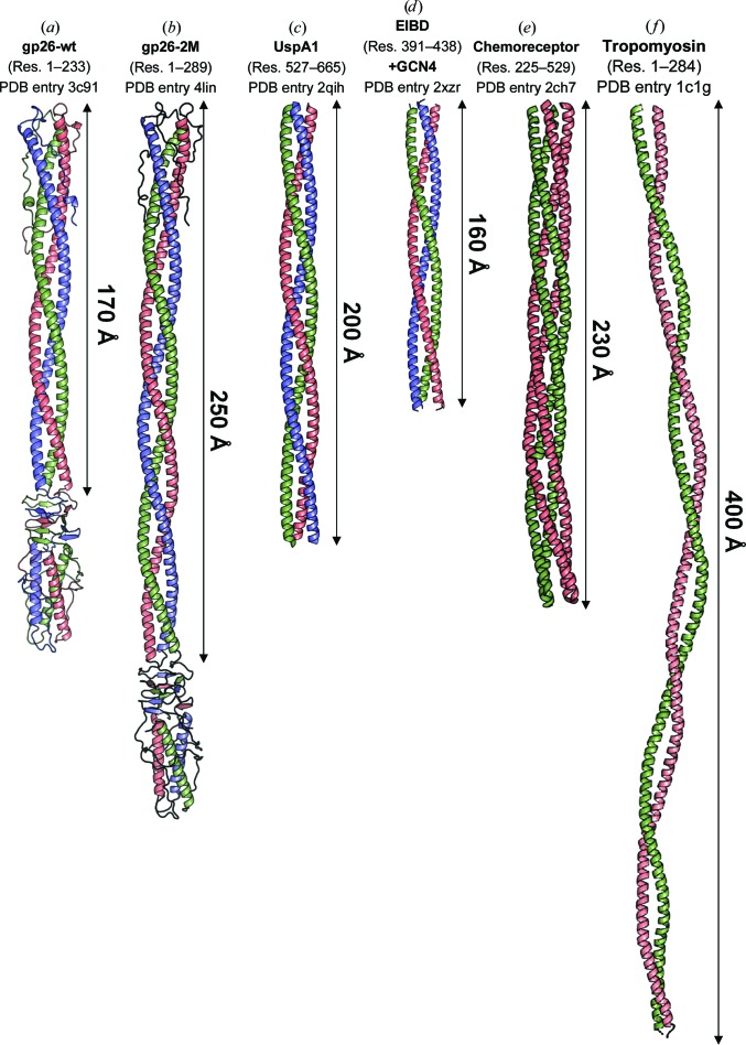
Figure 6.
Crystal structures of helical coiled-coil fibers longer than 100 Å. (a–d) The crystal structures of trimeric gp26-wt, gp26-2M, UspA1 (residues 527–665) and E. coli immunoglobulin-binding protein (EIBD) residues 391–438 fused to GCN4 adaptors (PDB entries 3c9i, 4lin, 2qih and 2xzr). (e) A 2.5 Å resolution crystal structure of the dimeric (tetrameric coiled coil) cytoplasmic domain of a bacterial chemoreceptor from T. maritima (PDB entry 2ch7). (f) A 7.0 Å resolution structure of tropomyosin (PDB entry 1c1g). The relative length of continuous helical regions is indicated.

