
Figure 1. Detection of γH2AX nuclear foci in MM patient samples and HMCLs.
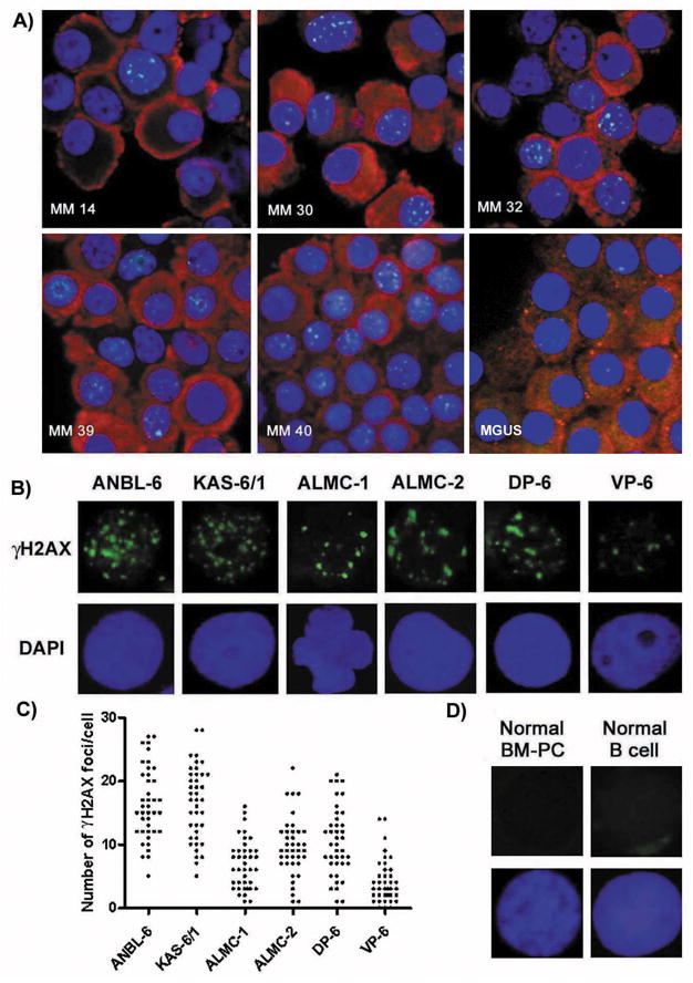
A) IF detection of γH2AX nuclear foci in freshly isolated CD138+ cells of 5 MM patients and 1 MGUS patient using an antiγH2AX-FITC Ab (green), anti-Ig (H + L) Ab (red) and DAPI (blue). B) Representative IF detection of nuclear γH2AX foci in HMCLs using an anti-γH2AX-FITC Ab (green) and DAPI (blue). C) The number of γH2AX foci/cell present in 50 individual cells for each HMCL. D) Representative IF detection of nuclear γH2AX foci in normal BM-PCs and normal B cells using an anti-γH2AX-FITC Ab (green) and DAPI (blue).