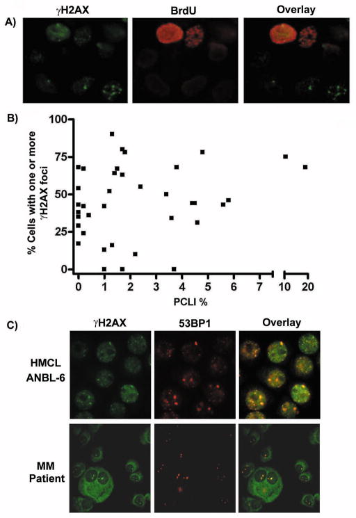
Figure 2. γH2AX foci do not correlate with phase of the cell cycle and γH2AX colocalizes with 53BP1.
A) IF analysis of BrdU labeled ANBL-6 cells revealed both BrdU positive (S/G2M phase) and BrdU negative (G1) cells possessing γH2AX foci. B) No correlation is observed between the % of cells possessing γH2AX foci in primary MM patient samples and their respective PCLI. C) HMCLs and primary MM patient samples were double stained with an anti-γH2AX-FITC Ab (green) and an anti-53BP1/Alexa 594 Ab (red). The resulting images were then overlaid in order to determine colocalization. Representative results are shown using one MM patient sample as well as the HMCL ANBL-6.

