
Figure 5.
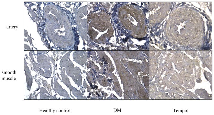
Immunohistochemistry of anti-nitrotyrosine. Nitrotyrosine protein expression in artery wall and smooth muscle cells of rat crura was higher in diabetic rats than in healthy controls. Tempol treatment decreased nitrotyrosine formation. Magnification is x200.