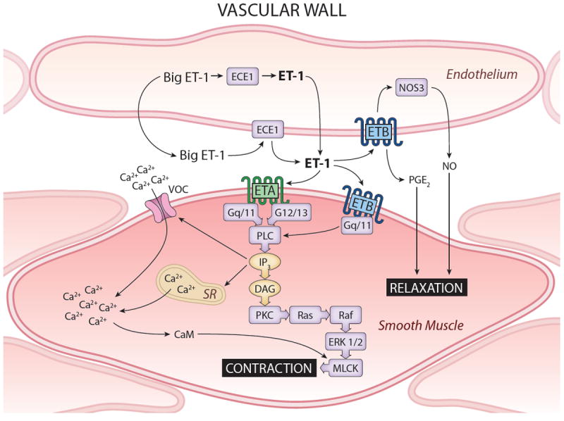
Figure 2.

Schema of ET in vasculature. Endothelial cells express ETB exclusively and are the predominant vascular source of ET-1. ET-1 and nitric oxide synthase 3 (NOS3) can increase ETB activity or amount, respectively, leading to NO and PGE2 production with resulting vasolaxation. Activation of vascular smooth muscle ETA or ETB leads to a signaling cascade involving G-proteins, phospholipase C (PLC) and inositol triphosphate (IP3) that activate voltage-operated Ca2+ channel (VOC) and sarcoplasmic reticulum (SR)-mediated increases in [Ca2+]i and calmodulin (CaM) activation. CaM, together with activation of protein kinase C (PKC) by diacylglycerol (DAG) and Ras/Raf/ERK1/2 activation, causes myosin light chain kinase (MLCK) activation and cell contraction.
