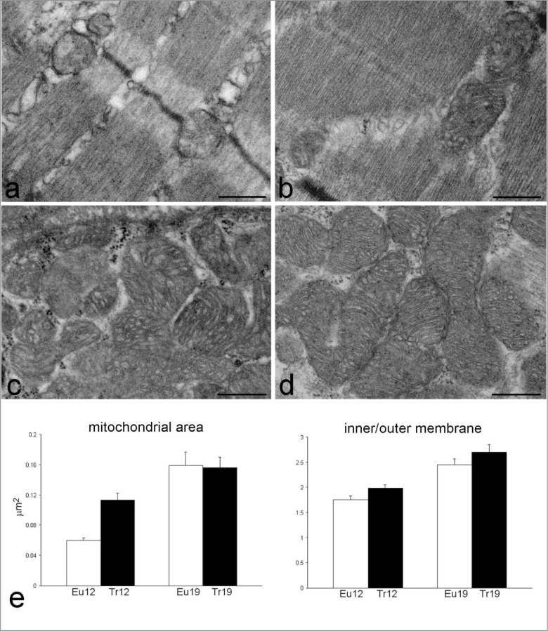
Figure 3.
Representative transmission electron micrographs of myofibres from adult euploid (a), adult trisomic (b), aging euploid (c) and aging trisomic (d) mice. Bars: 500 nm. (e) Results of morphometrical analysis on mitochondria: mitochondrial area and inner/outer membrane ratio (mean±SE) in 12- and 19 month-old euploid (Eu) and trisomic (Tr) mice. Two-way ANOVA revealed significant effect of the factors “trisomy” (P=0.043), “age” (P<0.001) and their interaction term (P=0.035) on the mitochondrial area; for the inner/outer membrane ratio significant effects were found for the factor “trisomy” (P=0.024) and “age” (P<0.001).

