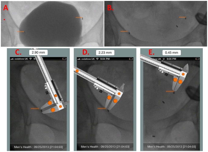
Figure 6. In order to confirm that the fiducial markers move apart with bladder filling and move together with bladder emptying, motion, immediately after all markers were placed, we performed a volumetric filling/emptying cystogram.
Using diluted contrast, the bladder was serially filled 0-arm remained fixed in position. The same procedure was repeated, separately, with saline. At each incremental 60 ml. change in bladder volume, we obtained a spot-fluoro image. We compared images of a patient's bladder filled with equal volumes of dilute contrast (A) and saline (B). We then used the digital image measurement App (MedMeasure!; U.S. and International Patents Pending) [https://itunes.apple.com/us/app/medmeasure!/id654898049?mt=8] to measure the distance between pairs of markers in paired images of the bladder filled with the same volume of saline or contrast. The digital, scalable caliper provided by the MedMeasure! App is first calibrated to one of the fiducial markers visible in lateral view– whose length of which is known to equal 2.9 mm. (C). Upon calibration, the actual distance between any two markers can be measured with the caliper. We measured the difference in distance between each marker-pair in paired images. (D, E).

