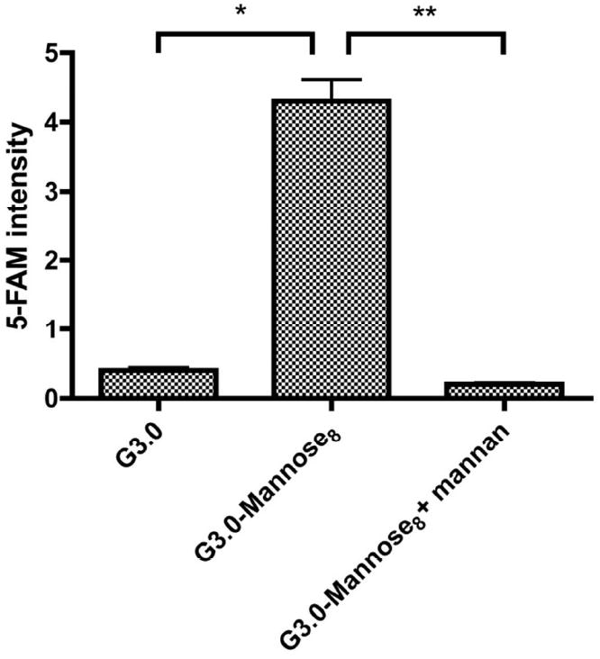
Figure 7.

Uptake of dendrons G3.0, G3.0-Mannose8 without mannan and G3.0-Mannose8 with mannan in J774.E murine macrophage-like cells. The uptake procedure was the same as described above in Fig.6. Uptake was quantified by 5-FAM green fluorescence of the confocal microscopy images with Leica software (LAS AF Lite, version 2.3.0). The values for G3.0, G3.0-Mannose8, and G3.0-Mannose8 with mannan were 0.4 ± 0.2, 4.3 ± 1.4, and 0.2 ± 0.1, respectively. Data is reported as mean ± standard deviation (n = 20 cells for each treatment, * P < 0.05, ** P < 0.05).
