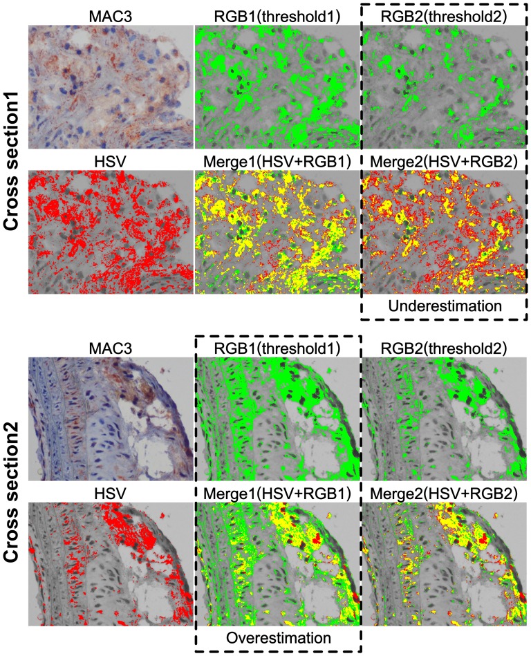
Figure 4. CSH is consistent between individuals.
Apoe−/− mouse innominate arteries stained with MAC3 antibody for detection of macrophages. RGB1(threshold1) was optimized for Cross Section 1, and overestimates the positive area when applied to Cross Section 2. RGB2(threshold2) was optimized for Cross Section 2, and underestimates the positive area when applied to Cross Section 1. CSH was able to effectively use a single HSV threshold on both cross sections. In the overlays between the HSV and RGB1 and RGB2, yellow area shows where there is agreement between the HSV method and the RGB method. Green area in the overlays may indicate false positive area reported by the RGB method, while red area in the overlays may represent false negative area reported by the RGB method.

