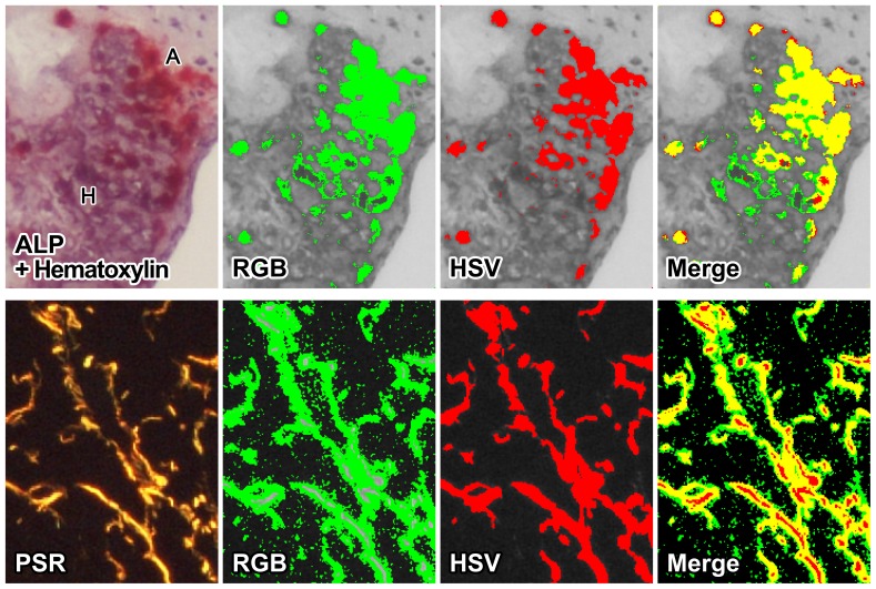
Figure 5. CSH is a powerful tool for a variety of stains.
Common histological stains, displaying the fidelity of CSH. (Top panels): A mouse aorta stained with alkaline phosphatase (ALP, red) for detection of early calcification, with Gill's hematoxylin as counterstaining (purple), which depicts advanced calcification. ALP stain is scarlet red (denoted “A” in the top left panel), while hematoxylin is a shade of purple (denoted “H” in the top left panel). Visually, the hematoxylin interferes with the ALP, making it difficult to see where the ALP stain begins and ends. We analyzed the section for ALP-positive area using both CSH and an RGB-based method. (Bottom panels): A mouse liver stained with picrosirius red staining visualized using polarized light microscopy for detection of fibrosis. We analyzed the section using both CSH and an RGB-based method. The RGB method was unable to register the brightest parts of the stain as positive (gray), and falsely interpreted stain artifacts as positive areas (green in both “Merge” images).

