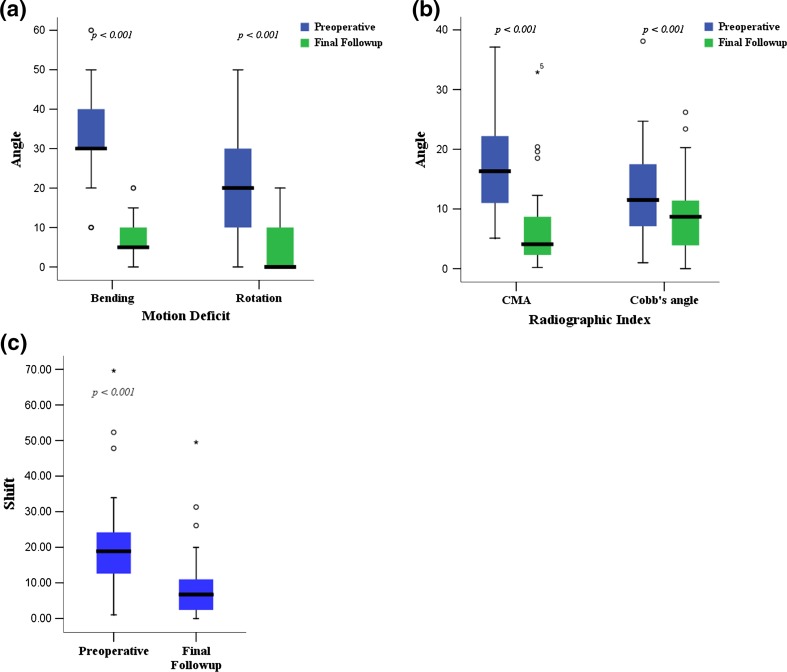
Fig. 3A–C.
The box plots show the comparisons of the parameters before surgery and at the latest followup. The (A) motion deficit was significantly reduced, and the (B) radiographic parameters such as cervicomandibular angle (CMA) and Cobb’s angle were significantly improved. (C) The lateral shift of head was significantly decreased at the latest followup.

