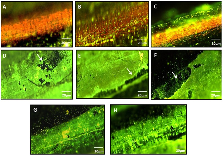
Figure 2.
A) Naked wire [not immersed in bacterial suspension] stained with fluorescent dye as viewed under 10x,Nikon eclipse 80i B) naked wire as seen with adhered bacteria (immersed for 6 h in bacterial susupension,107 CFU/ml) C) naked wire as seen with adhered bacteria (immersed for 24 h in bacterial susupension,107 CFU/ml) D) and E) Syto 9 stained HPMC coated wires (not immersed in bacterial suspension) showing coating on the wire F) and G) HPMC coated wires (immersed for 6 h in bacterial susupension,107 CFU/ml) as seen with adhered bacteria (seen as green spots) and H) HPMC coated wires as seen with adhered bacteria (immersed for 24 h in bacterial susupension,107 CFU/ml). (Note: Although the coating was uniform but the white arrows show slight discrepancy in the coating at some locations on wires)

