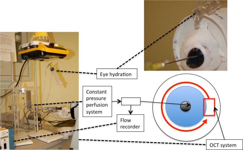
Figure 1. Experimental setting for ex-vivo imaging of anterior chamber outflow in porcine eyes.
A needle attached to a constant pressure perfusion system, whose flow is measured by the flow recorder, is inserted between the lens and iris in the posterior chamber. 12 OCT scans (red box) were taken along the limbus of the eye(red arrow). Dashed lines shows the equipment indicated by the boxes.

