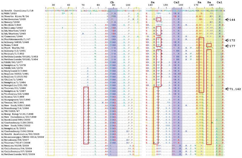
Fig. 1. Acquisition of glycosylation sites in HA of human H1N1 subtype over time prior to the emergence of the 2009 H1N1 virus.
Amino acid alignment of antigenic sites in the HA1 of seasonal H1N1 strains circulating in humans since 1918 until just prior to the emergence of the 2009 H1N1 pandemic virus. Four representative 2009 H1N1 pandemic isolates are also included as reference. Alignment shows selected prototypical reference strains. Colored shading (purple, yellow and green) depicts known antigenic sites listed on top. Yellow boxes represent conserved glycosylations and red boxes represent glycosylations that appear and disappear over time. Arrows on the right show the year glycosylations appeared at the indicated residue positions.

