
Figure 4. In a 42-year-old female with a 9×8 mm nodule on the left thyroid lobe, surgical pathology showed a subacute thyroiditis.
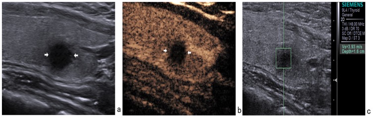
(a) Gray-scale US showed a hypoechoic nodule with ill-defined margin. (b) CEUS showed hypoenhancement. (c) The SWV value was 3.93 m/s when the region of interest was placed within the nodule. It was misdiagnosed as malignant nodule by conventional US, CEUS, and ARFI.