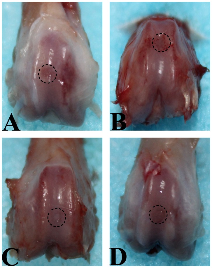
Figure 1. Macroscopic appearance of an operated knee at explantation after 10 weeks of follow-up.
A. SED group. Defect is partially filled with repair tissue. B. 2W group. The margin of the normal cartilage around defect was eroded. C. 4W group. Defect is completely filled with white cartilage-like repair tissue to the level of surrounding uninjured cartilage, and the junction area is obvious. D. 8W group. Defect is filled with a white cartilage-like tissue with an irregular surface, and there is transparent line in the boundary area between the repair tissue and the residual cartilage.

