
Figure 3. Histological photographs of the representative repair tissues of defects in four groups at 10 weeks after operation with safranin-O staining.
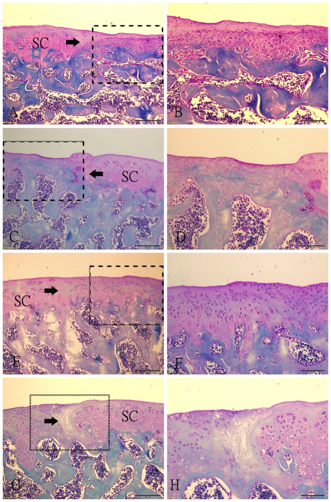
(A–B) Fibrous tissue in SED group. The spindle-shaped fibroblasts embeded in thin fibrin-like tissue are present in the tissue. (C–D) Fibrous tissue in 2W group. The regenerative fibrous tissue is much thinner than that in SED group, and there are a little fibroblasts. The margin of the normal cartilage adjacent the defect is destroyed. (E–F) Repair tissue in 4W group. A large number of rounded cells embedded in the intensely Safranin-O stained extracellular matrix. The integration of the lesion to adjacent articular cartilage is good. (G–H) Regenerative tissue in 8W group. Some clusters of rounded cells resembling chondrocytes were recognized embedded in a fibrous and extracellular matrix. The bonding area is acellular. Fig. B, D, F, H (scale bar = 200 µm) are higher magnifications of the junction of the repair tissue from Fig. A, C, E, G (scale bar = 500 µm) respectively. SC: surrounding cartilage. →: defect margin.