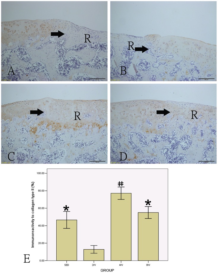
Figure 7. Immunohistochemical staining of collagen type II and the percentage of positive collagen type II in all groups (A: SED group; B: 2W group; C: 4W group; D: 8W group).
# P<0.05 compared to SED, 2W and 8W group; * P<0.05 compared to 2W group. (scale bar = 100 µm). R: repair tissue. →: defect margin.

