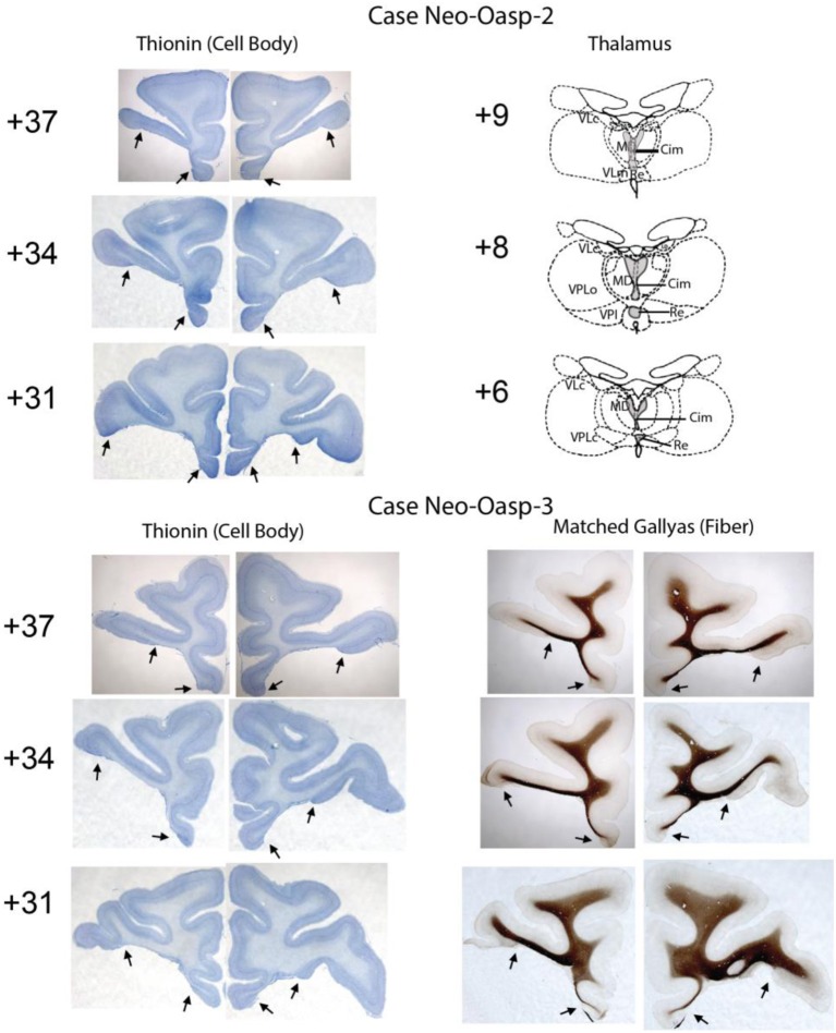
Figure 2.
Extent of neonatal OFC lesions is illustrated on thionin-stained sections for Cases Neo-Oasp-2 and -3 on the left column. Resulting thalamic degeneration following the neonatal OFC lesions is illustrated on drawings at three levels through the thalamus for a Case Neo-Oasp-2 and sparing of fibers underlying the OFC lesions is illustrated on Gallyas-stained sections for Case Neo-Oasp-3. Abbreviations: Cim, central intermedial; MD, mediodorsal; Re, reuniens; VLc, ventral lateral caudal part; VLm, ventral lateral, medial part; VPL, ventral posterior, lateral part; VPLo, ventral posterior, lateral oral part.

