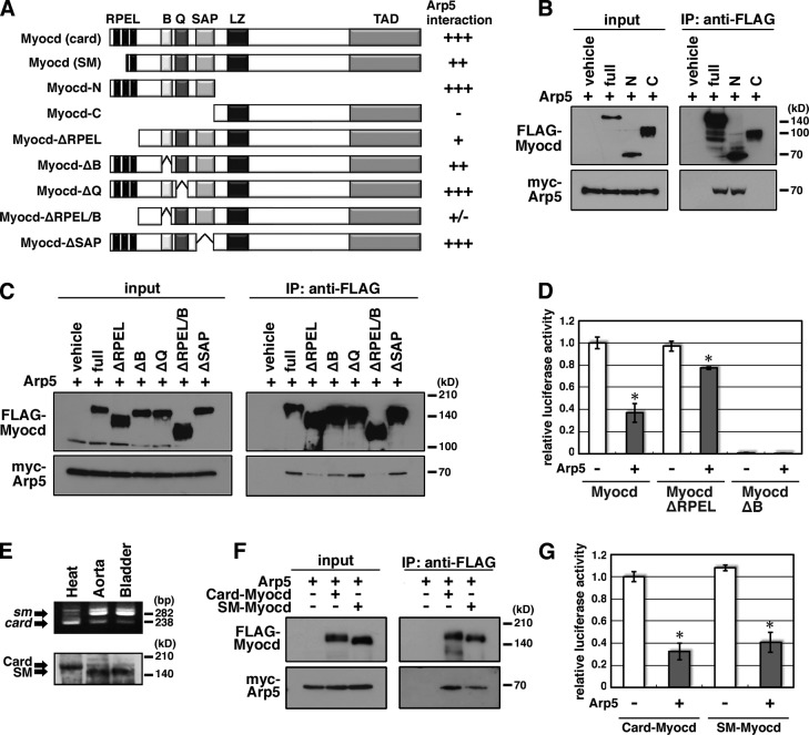
Figure 3.
Mapping the interaction domain of Myocd with Arp5. (A) Schematic representation of the domain deletion constructs of Myocd. RPEL, RPEL domain; B, basic region; Q, Q-rich region; SAP, SAP domain; LZ, leucine zipper; TAD, transcription activation domain. (B and C) Hek293T cells were transfected with the indicated combinations of expression vectors and coimmunoprecipitation was performed. (D) The indicated expression vectors were transfected into HeLa cells with 3× CArG-Luc, and the luciferase reporter assay was performed. Data represent the mean ± SEM of four independent experiments. *, P < 0.05, Student’s t test. (E) Expression pattern of SM/Card-Myocd in the rat heart, aorta, and bladder was determined by RT-PCR (top) and Western blotting (bottom). RT-PCR was performed with the primer pair for amplifying myocd exon 2–5, including (SM-type, top arrow) or excluding (Card-type, bottom arrow) exon 2a. In Western blotting, anti-Myocd antibody recognized high molecular weight Card-Myocd (top arrow) and low molecular weight SM-Myocd (bottom arrow). (F) Hek293T cells were transfected with the indicated combinations of expression vectors and coimmunoprecipitation was performed. (G) The indicated expression vectors were transfected into HeLa cells with 3× CArG-Luc, and the luciferase reporter assay was performed. Data represent the mean ± SEM of four independent experiments. *, P < 0.05, Student’s t test.

