Figure 7.
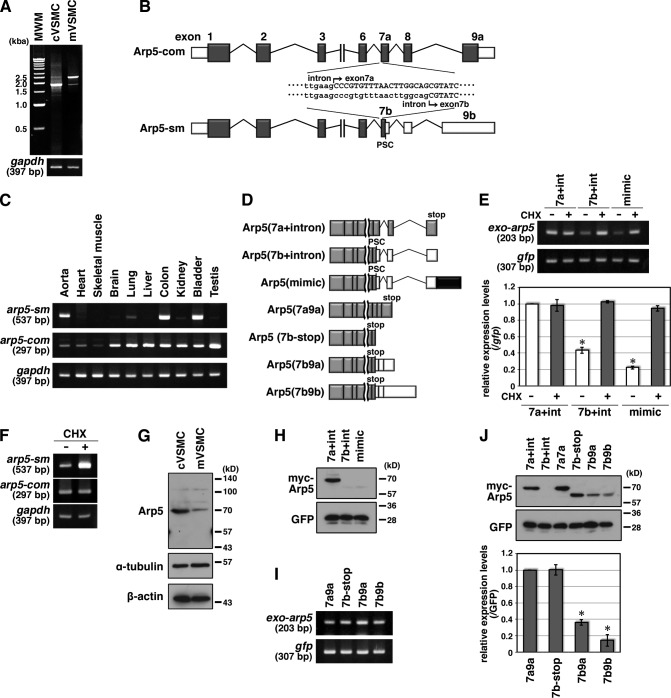
Quantitative regulation of Arp5 in smooth muscle cells by alternative splicing. (A) Total RNA was extracted from cultured VSMCs (cVSMC) and medial VSMC layers of abdominal aortae (mVSMC), and RT-PCR was performed with a primer pair for amplifying full-length arp5 mRNA. (B) Schematic representation of the alternative splicing of rat arp5. White box, 5′ and 3′UTR; gray box, exon; bar, intron. (C) Tissue-expression pattern of arp5-sm and arp5-com mRNA. Total RNA was extracted from the indicated rat tissues, and RT-PCR was performed with arp5 variant-specific primer pairs. (D) Schematic representation of expression constructs for arp5 variants. White box, 3′UTR; gray box, exon; black box, exon 9b characteristic sequence; bar, intron. (E) NMD of exogenous arp5 variants. HeLa cells were cotransfected with the indicated Arp5 variants and GFP expression vectors and treated with 100 µg/ml CHX or DMSO as a vehicle control for 4 h. Total RNA was extracted and RT-PCR was performed with a primer pair for amplifying exogenous arp5 (exo-arp5) or gfp (top panels). The amounts of PCR products were quantified by densitometry with normalization to gfp mRNA and statistically analyzed (bottom). Data represent the mean ± SEM of four independent experiments.*, P < 0.05, Student’s t test. (F) NMD of endogenous arp5 mRNA in cultured VSMCs. Total RNA was extracted from cultured VSMCs treated with CHX or DMSO for 4 h. RT-PCR was performed with arp5 variant-specific primer pairs. (G) Total proteins were extracted from cultured VSMCs (cVSMC) and medial VSMC layers of abdominal aortae (mVSMC), and Western blotting was performed with anti-Arp5 antibody and anti–α-tubulin and anti–β-actin antibodies as loading controls. (H) HeLa cells were cotransfected with the indicated Arp5 variant and GFP expression vectors, and Western blotting was performed with anti-myc and anti-GFP antibodies. (I) HeLa cells were cotransfected with the indicated Arp5 variant and GFP expression vectors. Total RNA was extracted and RT-PCR was performed with the indicated gene-specific primer pairs. (J) HeLa cells were cotransfected with the indicated Arp5 variant and GFP expression vectors, and Western blotting was performed with anti-myc and anti-GFP antibodies (top panels). The amounts of Arp5 variant proteins were quantified by densitometry with normalization to the GFP protein and statistically analyzed (bottom). Data represent the mean ± SEM of three independent experiments. *, P < 0.05, Student’s t test.

