
Figure 3.
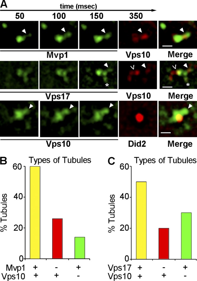
Retromer- and Mvp1-coated SNX-BAR–coated tubules contain retrograde cargo. (A) Galleries of representative time-lapse micrographs showing endosomes from cells coexpressing Vps10-2xmRFP and either Mvp1-GFP or Vps17-GFP and cells coexpressing Vps10-2xGFP with Did2-mKate2. A single tubule colocalized for both Mvp1 and Vps10 is shown in the top row. The middle row shows a Vps17-mGFP–decorated endosome and three types of tubules; a tubule decorated by both Vps17-GFP and Vps10-2xmRFP (filled arrowhead), a tubule containing Vps10-2xmRFP but not Mvp1-GFP (<), and a tubule containing Vps10-2xmRFP but not Vps17-GFP (*). The bottom row shows time-lapse micrographs of a Did2-mKate2 endosome from which a tubule containing Vps10-GFP, but not decorated by Did2-mKate2, is budding (arrowhead). The end point (milliseconds) of each exposure is indicated on the top. Bar, 500 nm. (B) The proportion of tubules decorated by Vps17-GFP and containing Vps10-2xmRFP in wild-type and mvp1Δ cells is plotted. (C) The proportion of tubules in cells expressing Mvp1-GFP and Vps10-2xmRFP is plotted.