
Figure 1.
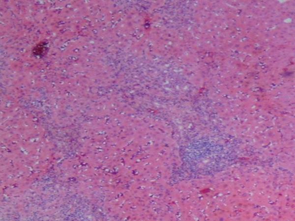
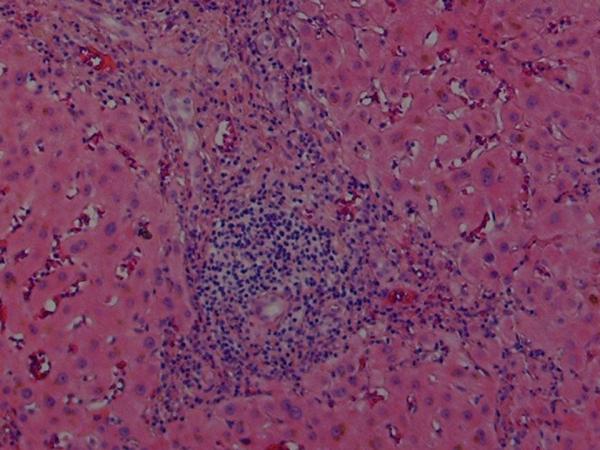
Histologic findings in the liver of a patient with celiac disease and hypertransaminasemia; liver tests returned to normal after 6 months of strict adherence to a gluten-free diet, no follow-up liver biopsy was seem necessary
(A) Low-magnification showing periportal inflammation and mononuclear infiltration on the parenchyma (hematoxylin and eosin)
(B) High-magnification showing the portal triad with extensive mononuclear infiltration. Note the sparing of the biliary ducts (hematoxylin and eosin)