Figure 2.

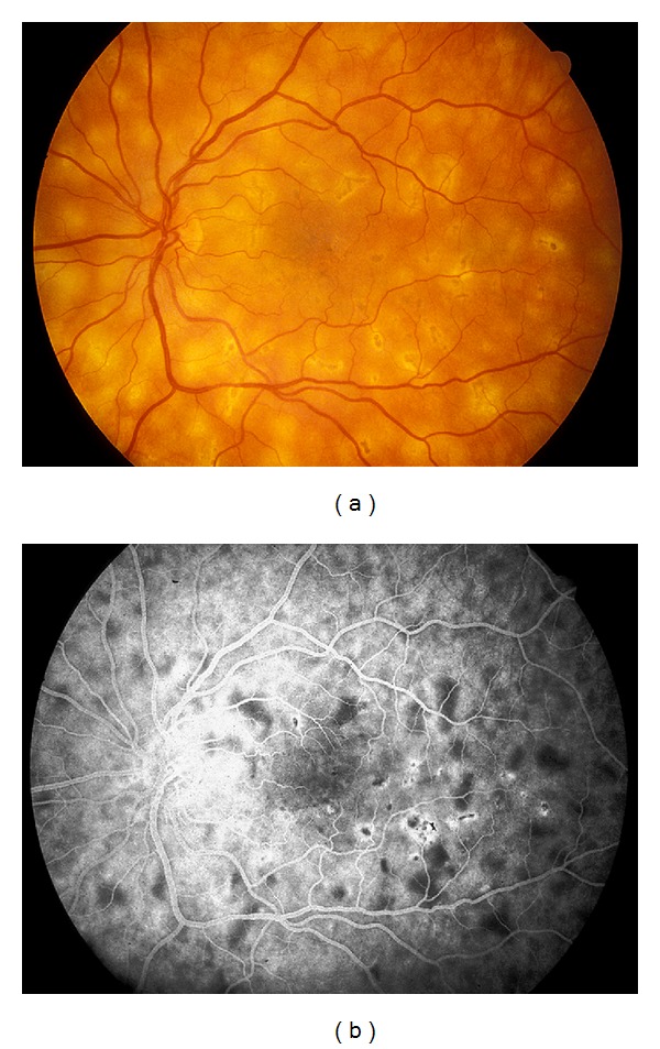
(a) Fundus photograph and corresponding (b) midphase fluorescein angiogram showing blockage of some lesions and the beginning of staining of other lesions as the later phase begins in APMPPE.

(a) Fundus photograph and corresponding (b) midphase fluorescein angiogram showing blockage of some lesions and the beginning of staining of other lesions as the later phase begins in APMPPE.