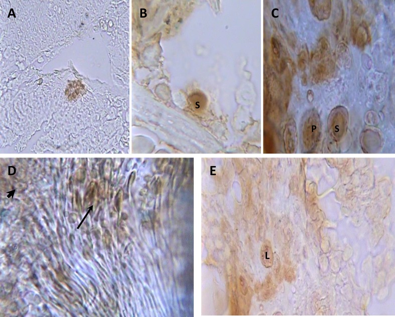
Figure 3.
Photomicrograph of longitudinal sections of testes in BrdU labeled MSCs injected group after monoclonal anti BrdU staining and alcian blue counterstaining. Colony of MSCs (A) in seminiferous tubules (400×); spermatogonium (S) (B) and spermatogonia (S) and primary spermatocytes (P) (C), sperm cluster (thin arrow) and spermatids (thick arrow) (D) in seminiferous tubules (1000×) and (E) Leydig cells (L) between seminiferous tubules (1000×).

