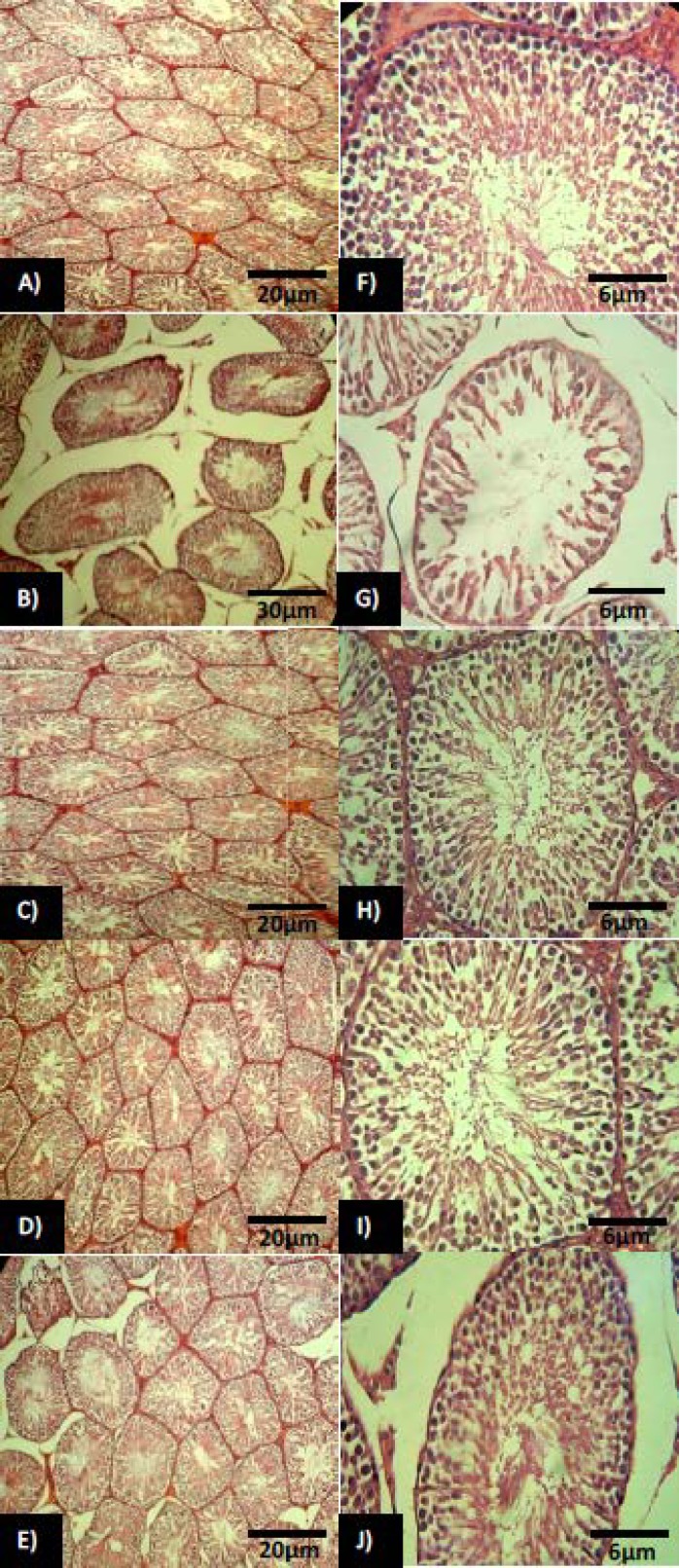
Figure 1.
Cross section from testis; (A) control group: note normal testicular tissue with normal seminiferous tubules in higher magnification (F). (B) Non-treated diabetic testis: the seminiferous tubules are degenerated and remarkable edema is manifested in interstitial tissue. Note negative TDI and SPI in higher magnification (G). (C) Metformin and honey co-treated group, showing normal spermatogenesis (H) with decreased edema. (D) Honey alone-treated group: The tubules are appeared approximately normal accomplished with significant decrease in edema. Note the normal spermatogenesis in higher magnification (I). (E) Metformin alone-treated group: The tubules are presented with higher thickness of germinal epithelium (J) while the mild edema remained in interstitial tissue. H&E staining technique, (A, B, C, D, E: 100× and F, G, H, I, J: 400×).

