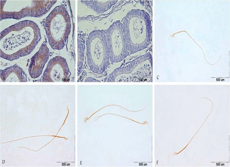
Figure 3.
(a) Strong immunoreactivity of CatSper 2 was detected in the lumen of epididymis in the young control group. (b) Epididymis section of young control group without primary antibody, CatSper1, no reaction was observed. (c,d) localization of CatSper 1 protein in the head and sperm tail of the control group 1 (c) and experimental group 1 (d). (e, f) localization of CatSper 1 protein in the head and sperm tail of the control group 2 (e) and experimental group 2 (f). In all slides, immunoreactions visualization was done with DAB and counterstained with haematoxylin. Scale bars represent 500 µm.

