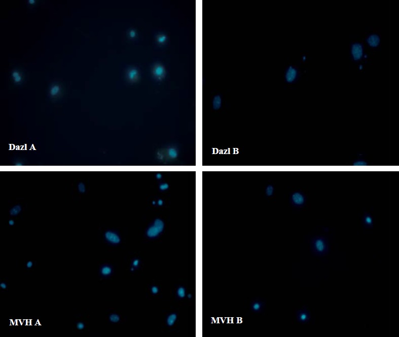
Figure 5.

Imunoflorescence staining of Mvh and Dazl (green) in bone marrow mesenchymal stem cells (a) and adipose tissue mesenchymal stem cells (b) [the group cutured in a medium without differentiating factor]. The nucleus were stained with DAPI (blue). Both kinds of the cells did not express Dazl and Mvh. [X400 magnification] .
