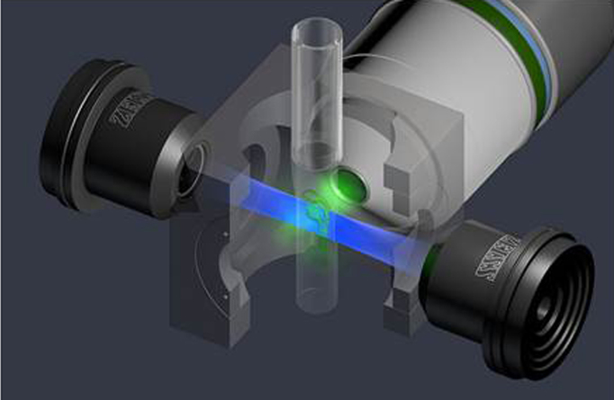
Figure 6.
Single plane illumination microscopy (SPIM) light path in the Zeiss Lightsheet Z.1 microscope (light sheet fluorescence microscopy: LSFM) using a whole zebrafish embryo. Illumination lenses (black), detection objective: W Plan-APHCHORMAT 20x/1.0 (white), lightsheet (blue). LSFM separates fluorescence excitation and detection into two separate light paths, with the axis of sheet illumination orthogonal to the detection axis (green). Laser light is focused into a thin light sheet by cylindrical lenses (not shown) upstream of the illuminating objectives. Only the in-focus light sheet excites fluorescence, significantly reducing phototoxicity. Use of two oppositional illuminating objectives reduces shadowing artifacts and increases acquisition speed. Fluorescence is collected on ultrafast CMOS cameras enabling z-stack acquisition speeds >100-fold faster than laser scanning confocal microscopy. Image courtesy of Carl Zeiss Microscopy GmbH. Used with permission.

