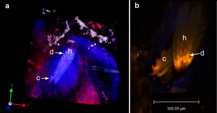
Figure 4.

Snapshots of two photon images of the mouthparts after insertion into the dermis. a) Snapshot of Z-stack movie of the skin bite site at 72 hours of tick attachment. The 3-dimensional z-stack image was rendered using Volocity software from images of sequential xy planes taken at different levels in the tissue. Note the dense cone-shaped appearance of the blue collagen fibers surrounding the hypostome. These fibers appear blue due to second harmonic generation. Rhodamine-conjugated dextran was injected intravenously prior to imaging to demonstrate the extravasation of blood adjacent to the hypostome. b) Ventral aspect of the hypostome with a single chelicera extended. h = hypostome, c = chelicera, d = denticle.
