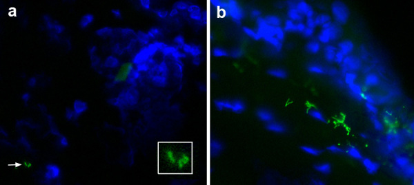
Figure 6.

Direct immunofluorescence of B. burgdorferi in ear skin after tick feeding. a) Rare spirochete forms (green) are seen in the dermis (arrow) at day 5 after tick attachment (enlarged in insert). Skin cell nuclei (blue) are stained with DAPI. The tick had spontaneously detached prior to this analysis. b) Spirochetes are more numerous at day 7 after tick attachment (the tick had detached on day 4).
