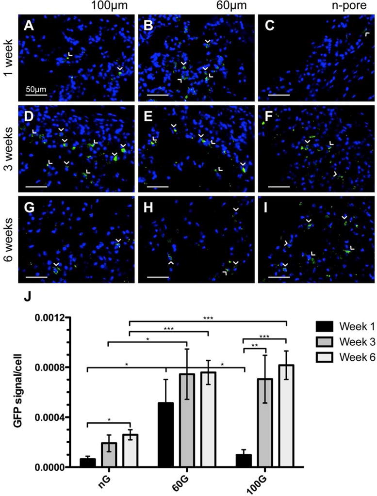
Figure 3.

Immunofluorescence staining of GFP in pGFPluc loaded control 100 (A, D, G) and 60 μm (B, E, H) porous and n-pore (C, F, I) hydrogels at 1 (A, B, C), 3 (D, E, F), and 6 (G, H, I) weeks indicates several transfected cells are present in each hydrogel over the 6 week period. Transfected cells in n-pore hydrogels, however, are only located around the hydrogel periphery where there is infiltration and gel degradation, while transfected cells in 100 and 60 μm porous gels can be found throughout. (J) Quantification of GFP positive cells normalized to total cells per image area reveals statistical differences in transfection levels at all times between 60 and 100 μm porous and n-pore hydrogels. GFP positive cells = green, cell nuclei = blue. All images are 40× magnifications.
