Figure 4.

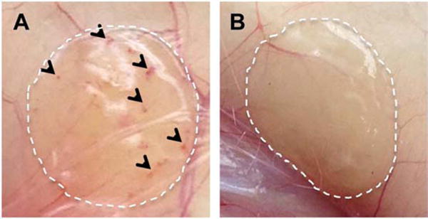
Digital images of pVEGF loaded 60 μm porous (A) and n-pore (B) hydrogel implants upon excision at 6 weeks demonstrate visible differences in angiogenesis.

Digital images of pVEGF loaded 60 μm porous (A) and n-pore (B) hydrogel implants upon excision at 6 weeks demonstrate visible differences in angiogenesis.