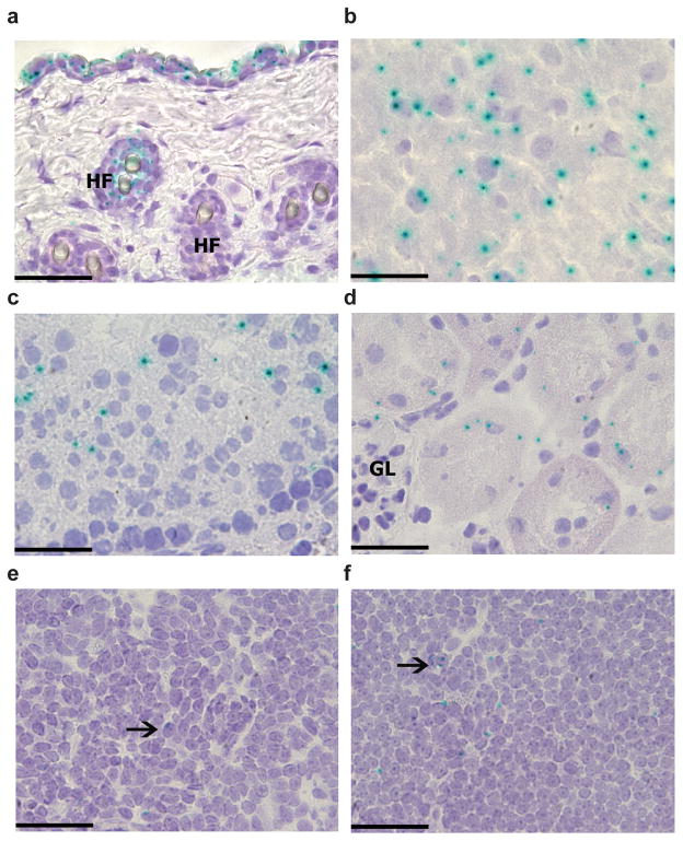
Figure 1.
Tissue specific expression of IL-34. Staining for X-gal (blue) in skin (a), brain cerebral cortex (b), testis (c), kidney tubules (d), spleen (e) and lymph nodes (f) from Il34LacZ/LacZ mice. Tissues from wild-type mice did not show any X-gal staining (data not shown). HF, hair follicles; GL, kidney glomerulus. Arrows indicate X-gal+ cells. Original magnification, ×400 (a; scale bar, 50 μm) or ×600 (b–f; scale bars, 33 μm). Data are representative of 2 experiments with three mice (25 high-power fields per tissue per mouse).

