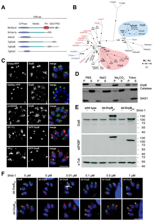
Figure 1. DrpB belongs to a ciliate specific class and localises close to the Golgi.
(A) Scheme of different representative of the dynamin family, including T. gondii DrpA,B,C. Three representatives of dynamin proteins are shown considering their domain architecture (Sc: Saccharomyces cerevisiae; Mm: Mus musculus; At: Arabidopsis thaliana). (B) Phylogenetic analysis of DrpA and DrpB reveals that they belong to unrelated classes. The figure shows an unrooted Maximum Likelihood phylogenetic tree (Ln Likelihood = −33260.47) in which bootstrap values >50% based on Maximum Likelihood and Distance Matrix/Neighbor-Joining phylogenies are indicated on the respective branches. Dynamins required for endocytosis are indicated in blue and required for mitochondria division in red. DrpA and DrpB fall into two distinct clades (indicated in purple and blue respectively). The accession numbers and the alignment can be downloaded as supporting information (Bb: Babesia bovis; Ce: Caenorhabditis elegans; Cm: Cyanidioschyzon merolae; Cp: Cryptosporidium parvum; Cr: Chlamydomonas reinhardtii; Dm: Drosophila melanogaster; Hs:Homo sapiens; Lm:Leishmania major; Os:Oryza sativa; Pf:Plasmodium falciparum; Pr: Phytophthora ramorum; Ps: Phytophthora sojae; Pte: Paramecium tetraurelia; Pv: Plasmodium vivax; Sp: Schizosaccharomyces pombe; Ta: Theileria annulata; Tb: Trypanosoma brucei; Tg:Toxoplasma gondii; Tp: Thalassiosira pseudonana; Tt: Tetrahymena thermophila) (C) Immunofluorescence analysis of RHWT parasites grown on HFF-monolayers, using the indicated antibodies (proM2AP and VP-1) or transiently expressing the indicated markers (GRASP-RFP, GalNac-YFP and AP-1Ty, GFP-DrpB).. Scale bar: 10 μm (D) Cell fractionation on wild type parasites. Extracellular parasites were harvested and lysed under different conditions (PBS, PBS with 1 M NaCl, PBS with 0.1 M Na2CO3 (pH 11.5) and PBS with 2% Triton X-100) followed by ultracentrifugation. Supernatant (SN) and pellet (P) fractions were analyzed by western blot analysis with indicated antibodies. (E) Immunoblot analysis of clonal parasites transfected with dd-DrpBWT or dd-DrpBDN. Both parasite strains express the respective fusion protein only in presence of Shld-1. The immunoblot was probed with the indicated antibodies. TUB1 served as a loading control. (F) DrpBwt and DrpBDN accumulate close to the Golgi even at low overexpression. Parasites expressing GRASP-RFP and dd-DrpBwt or dd-DrpBDN were treated for 3 hours with the indicated amount of Shld-1 before localisation of GRASP and DrpB was compared. In case of dd-DrpBwt a signal for GFP can detected even at 0.01 μM Shld-1 close to the Golgi (arrow). Left: parasites not treated with Shld-1 were analysed using DrpB-antibodies. Scale bar: 10 μm. DrpB is always shown in green colour in merged images.

