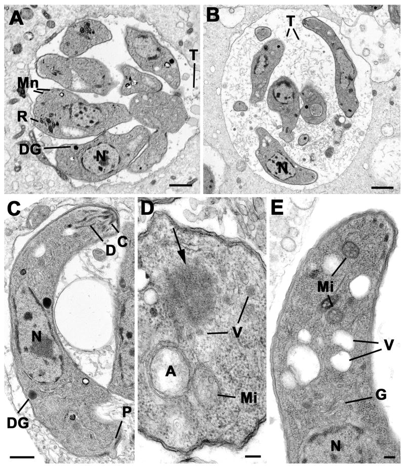
Figure 6. Transmission electron micrographs of control and treated intracellular parasites.
(A) Low power of a group of tachyzoites from the untreated control sample at 36 hours showing the parasites located within a relatively tight PV with few intra-vacuolar tubules (T). The tachyzoites have a central nucleus (N) and apical organelles consisting of rhoptries (R), micronemes (Mn) and dense granules (DG). Scale Bar is 1 μm. (B) Low power from at treated sample (36 hours) showing the parasites located within a loose PV with numerous intra-vacuolar tubules (T). Note the elongated appearance of the tachyzoites and the lack of apical organelles. N- nucleus. Bar is 1 μm. (C) Longitudinal section through a Shld-1 treated tachyzoites showing the centrally located nucleus (N), the conoid (C) and associated duct-like structures (D). Note the absence of rhotries and micronemes. DG – dense granule; P – posterior pore. Bar is 500 nm. (D) Cross-section through the anterior cytoplasm of a Shld-1 treated tachyzoite in which the apicoplast (A) and mitochondrion (Mi) can be seen. Note the homogenous electron dense structure (arrow) with surrounding vesicles (V). Bar is 100 nm. (E) Longitudinal section through the anterior of a Shld-1 treated tachyzoite illustrating a number of electron lucent vacuoles (V) located anterior to the Golgi body (G). N – nucleus; Mi – mitochondrion. Bar is 100 nm.

