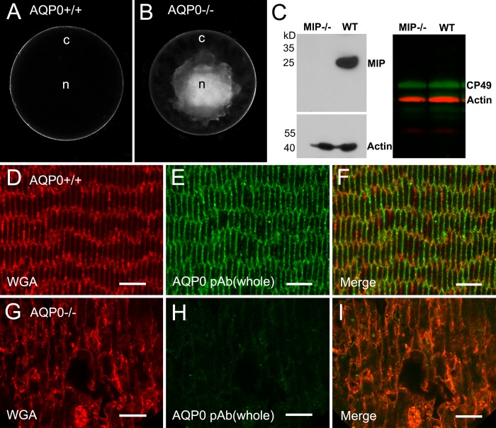
Figure 1.
Formation of nuclear cataract and loss of AQP0 protein in an AQP0-deficient lens. (A, B) The wild-type control displays a whole transparent lens compared with the appearance of dense cataract in the lens core in the AQP0−/− lens at age 5 weeks. (C) The absence of AQP0 proteins in AQP0−/− fiber cell membrane preparations is determined by Western blot (left). The presence of the CP49 beaded filament proteins is detected in both AQP0−/− and wild-type lenses with Western blot (right). By confocal immunofluorescence, labeling of AQP0 polyclonal antibody is present in cortical fiber cell membranes in the wild-type control (D–F), but is absent in the AQP0−/− cortical fibers (G–I). Note that swollen fiber cells are shown in the AQP0−/− lens. Rhodamine-conjugated wheat germ agglutinin was used to delineate the fiber cell membrane outlines. Scale bars: 5 μm.

