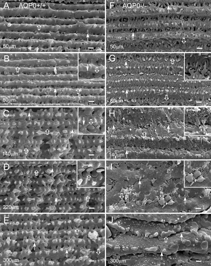
Figure 3.
Interlocking protrusions exhibit dramatic structural changes as fiber cells mature in an AQP0−/− lens. Structural comparisons of protrusions are made from the narrow-side fiber cells in different cortical regions between the wild type and AQP0−/− at age 4 weeks. (A–E) In wild-type lenses, interlocking protrusions (50–300 μm deep from the equatorial surface) undergo various stages of development and maturation. Many small protrusions are seen at approximately 50 μm in the superficial cortex (A). They grow and mature into unique configurations in deeper mature cortical fibers (B–E). Typically, two different configurations of protrusions are identified based on their unique shapes (see the insets in [B–D]). Type 1 normally consists of a narrowed neck and expanded head portions, with the head diameter in the range of 0.5 to 2 μm (open arrows). Type 2 generally displays a slender, tubular shape, with a diameter of approximately 0.1 to 0.3 μm (arrows). (F–J) In an AQP0−/− lens, protrusions of both types undergo similar stages of formation and growth but show signs of minor abnormalities at approximately 50 μm deep in the absence of AQP0 (F). As fiber cells mature, these protrusions surprisingly undergo uncontrolled elongation ([G, H] and insets), deformation (insets in [H, I]), and breakdown (I, J). Severe deformation of the protrusions eventually results in separation and collapse of fiber cells (I, J). Scale bars: 1 μm.

