Figure 5.
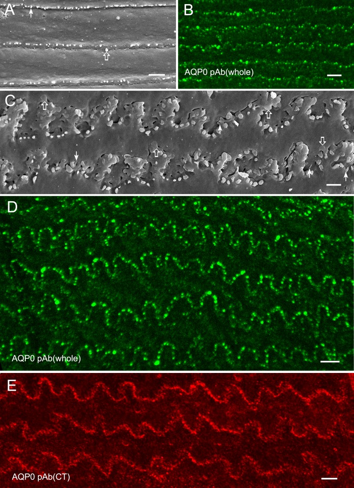
Aquaporin-0 is particularly enriched in protrusions examined with confocal immunofluorescence. (A) Scanning electron microscopy shows that in superficial fibers type 1 (open arrows) and type 2 (arrows) small protrusions are distributed along fairly straight broadside fiber cells in whole-mount preparations of wild-type lenses. (B) Immunofluorescence exhibits a dotted pattern of labeling for AQP0 polyclonal antibody against the whole molecule along the broadside superficial fiber cells, which correlates well with the straight distribution of protrusions seen on SEM. (C) Scanning electron microscopy shows the distribution of type 1 (open arrows) and type 2 (arrows) protrusions along the undulating boarders of broadside fiber cells in the deeper cortex. (D) Immunofluorescence reveals the distinct undulating dotted pattern of labeling for AQP0 polyclonal antibody against the whole molecule, which correlates well with the similar undulating distribution of protrusions along the broadside fiber cells in the deeper cortex seen on SEM. (E) Using an AQP0 C-terminal polyclonal antibody, its undulating dotted pattern of labeling is also consistent with the undulating distribution of protrusions along the broad side of deeper cortical fiber cells. Scale bars: 2 μm in (A, C); 5 μm in (B, D, E).

