Figure 6.
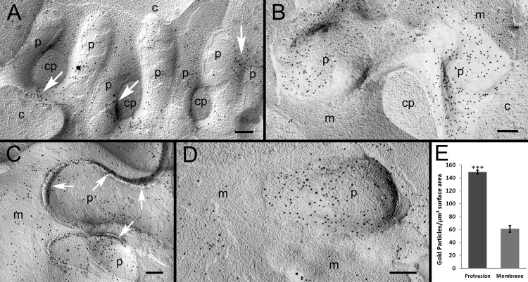
Preferential accumulation of AQP0 in protrusions examined with immunogold labeling. (A) The specific localization of AQP0 polyclonal antibody, represented by 10-nm gold particles, is observed on a cluster of protrusions (p). The rich accumulation of gold particles is distributed in the localized areas between protrusions, where they closely interlock (arrows). The high specificity of AQP0 labeling is evidenced in this figure, showing that the particles are localized mainly on the membrane of protrusions but not on the cytoplasm of protrusions (cp) and other cell cytoplasm backgrounds (c). (B) Representative labeling reveals that AQP0 is more densely labeled on protrusions (p) than the adjacent cell membranes (m). (C) The heavier labeling of AQP0 is also observed in the extracellular space (arrows) between protrusions. (D) In this study, quantitative analysis was conducted on micrographs (such as [D]) that contain both protrusions (p) and adjacent flat nonprotrusion membranes (m; see the Methods section). (E) A total of 5136 gold particles was counted, and the analysis showed that protrusions have significantly more gold particles than surrounding flat nonprotrusion membranes (*P < 0.001). Scale bars: 200 nm.

