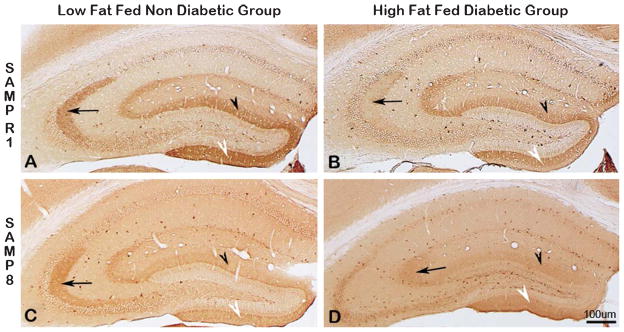
Fig. 9.

Immunodistribution of synaptophysin (SYN) in the hippocampus of SAMPR1 (A, B, top panel), and SAMP8 (C, D, bottom panel); low-fat fed non-diabetic mice (A, C, left panel), and with high-fat fed diabetic mice (B, D, right panel). Note the strongest SYN immunoreactivity (IR) within the CA3 dendritic field (Fig. 9A, arrow), and within the supra-granular (Fig. 9A, black arrowhead) and infragranular (Fig. 9A, white arrowhead) molecular layers of DG of non-diabetic SAMPR1 mice. Note that experimental induction of diabetes in SAMPR1 mice resulted in the reduction of SYN immunoreactivity only in the CA3 dendritic field (Fig. 9B, black arrowhead), while preserving SYN immunoreactivity within the supra-granular (Fig. 9B, black arrowhead) and infragranular (Fig. 9B, white arrowhead) molecular layers of DG of diabetic SAMPR1 mice. On the other hand, in non-diabetic SAMP8 mice, SYN immunoreactivity in the CA3 dendritic field (Fig. 9C, black arrowhead) was preserved, but the SYN immunoreactivity within the supra-granular (Fig. 9C, black arrowhead) and infragranular (Fig. 9C, white arrowhead) molecular layers of DG was reduced. In the hippocampus of diabetic SAMP8 mice, SYN immunoreactivity in all CA3 (Fig. 9D, arrow) and respective DG subfields (Fig. 9D, black arrowhead and white arrowhead) were observed to be greatly reduced. Scale bar = 100 μm.
