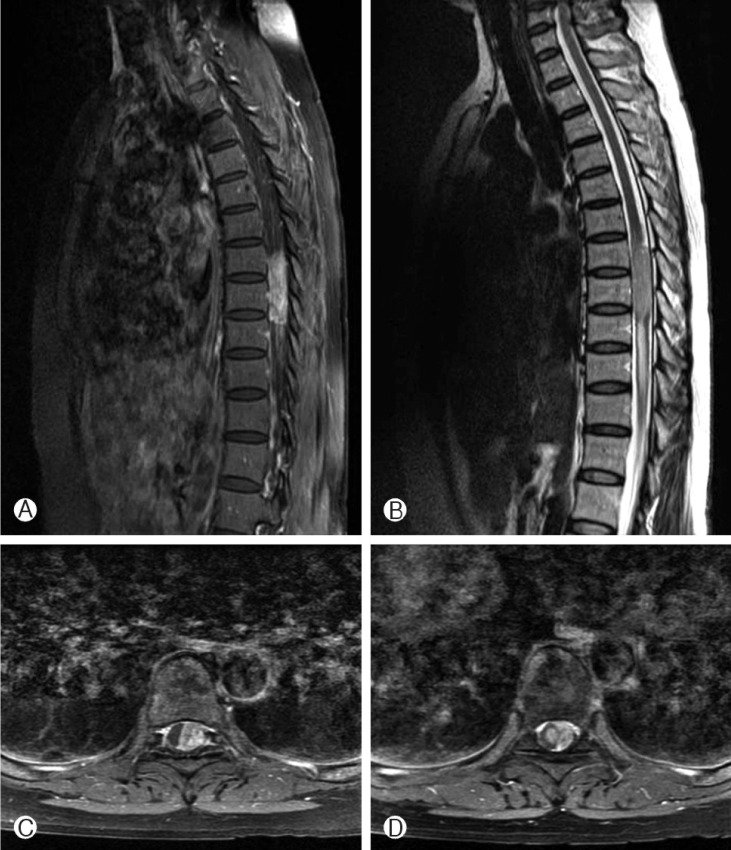
Fig. 1.
Preoperative spinal MRI. (A) T1-weighted sagittal MRI with the contrast showing an oval shaped enhanced mass at T7-9. (B) T2-weighted sagittal MRI showing a mass with slightly high signal intensity at T7-9 and cord signal change at T9-11. (C) T1-weighted axial MRI with the contrast showing an enhanced mass displacing the cord to right side at T8. (D) T1-weighted axial MRI with the contrast showing an enhanced mass displacing the cord to oppo site side and an enhanced small round shape mass within the cord is presented at T9.

