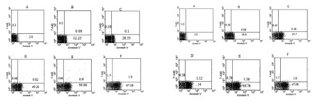
Figure 2. Dot plot view for induction of apoptosis with various concentrations of γ-T (0, 25, 50, 100 and 200μM) using annexin V– CF647/7-AAD staining and flow cytometry in HT29 cell line for 48h and 72h. CF647-labeled annexin V was an early marker for phosphatidyl serine externalization over cell membrane (lower right quadrant) and late apoptosis was labeled with 7-AAD in the upper left quadrant. HT29 cells were grown in DMEM, exposed to specific agents and early apoptosis was determined by annexin V staining method. Cells in positive control group received 2μg/ml/2h anisomycin. A) Untreated cells. B, C, D and E are groups that received 25, 50, 100 and 200μM γ-T. F) Treated cells with anisomycin 2μgr/ml/2h.